Let’s take a look at medical tattoos. For now, we’ll assume there are no data privacy/insurance equity concerns that would accompany such technology, and simply appreciate the biological aspect. Let’s take a look at tattoos that are meant for individual cells, heart tattoos, and a couple more.
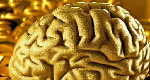
Gold Arrays for Brains
Starting with one that was just published1 recently: Imagine if each of your cells could be given a tattoo that would track their wellbeing. And furthermore, the tattoo would be made of gold.
Their process was nicely summarized2 by ScienceDaily as follows:
“The researchers built the tattoos in the form of arrays with gold, a material known for its ability to prevent signal loss or distortion in electronic wiring. They attached the arrays to cells that make and sustain tissue in the human body, called fibroblasts. The arrays were then treated with molecular glues and transferred onto the cells using an alginate hydrogel film, a gel-like laminate that can be dissolved after the gold adheres to the cell. The molecular glue on the array bonds to a film secreted by the cells called the extracellular matrix.”
Their work is notable because we’ve used hydrogels to adhere nanowires and nanodots on human skin before, but this is proof of concept for deliberate, careful patterns on cells.
They had two types of cell targets: the surface of rat brains, and coronal rat brain slices. Their gold arrays “selectively adhered to the surface of the whole rat brain”, but the wires on the slice were washed away after a rinse with EDTA. It is a chelating agent, meaning that it is able to break molecules away, acting as a sort of sticky hook. Otherwise, the arrays were able to remain on the moving cells for 16 hours.
This is a good start, but the arrays need to become more durable and display higher adhesiveness for longer-term applications. One interesting tidbit, while we’re here: the layers of gold are so thin that if we could see them, they would most likely be purple.
When we’re able to extend this to human applications, we would be able to monitor cell health to an incredible extent, allowing us to detect diseases in their early stages. The nanodots and nanowires would be arranged to create sensors and other components in the nanocircuit as necessary. More specifically, we can potentially design immunosensors that use antibodies to detect antigens, or enzyme-based biosensors that recognize specific substrates.
Art for your Heart
This cardiac implant made of graphene has all the capabilities that make it a pacemaker - yet its structure is light, thinner than a strand of hair, and flexible. This allows it to take shape to fit the shape of the heart.
Notably, it was tested for the treatment of atrioventricular block (a heart condition where electrical signals from the atria to the ventricles are interrupted) in a rat model.3 The graphene implant reported stable electrochemical measurements for 2 months, which fits short-term applications. The authors recommended that the next steps, especially when scaling this up to human hearts, include: accounting for tissue growth over the ultra-thin implant, more effective bioadhesives, additional studies on long-term toxicity, and mitigating the effects of additional adipose tissue on signals.
Designs for biomarker signs
One of my favourite papers is one that acts as a proof of concept for using tattoos to monitor and alert for various biomarkers and other indicators - it specifically focuses on blood calcium. Mildly elevated levels are an early symptom of many cancers, including breast, colon, and lung.4 This is significant - we may be able to detect cancer in its earlier stages, before most of the concerning symptoms surface. Early detection is greatly important to survival rates.
Here’s how the tattoo works: cells were designed with a specific calcium-sensing receptor, modified so that upon persistently increased blood calcium levels, would set off a chain of signals that would activate transgenic tyrosinase. This enzyme is in melanocytes, which, as implied, are specialized cells that produce melanin.5 The increased production of melanin produces a visible contrast that in the mouse models, was visible to the naked eye.
The researchers did a variety of RCTs, including one where they injected the tattoo into rats that had normocalcemic tumours, and another group that had hypercalcemia-associated tumours, positive controls, negative controls, etc.
Calcium is not the only biomarker we have a proof of concept for. Another study in 2021 showed successful detection of pH, uric acid, glucose and temperature.6 These all indicate potential health problems. For instance, high levels of uric acid occur when someone has kidney stones, and high blood sugar levels can help identify diabetes. Unlike the previous experiments mentioned above, this one was conducted on rabbit skin.
Future Applications
There’s much more to be done. We’ve had incredible research that demonstrates proof of concept, but that’s still a long way off from using these in the field. Biologically, we’re on a good track as we get closer to moving models to humans. Yet, the implications of the technology need to be addressed. How can we continue making these affordable? What would patient confidentiality look like? How would we ensure that there is informed consent around data privacy? How would we mitigate any potential stigmas?
https://pubs.acs.org/doi/10.1021/acs.nanolett.3c01960
https://www.sciencedaily.com/releases/2023/08/230807133828.htm
https://onlinelibrary.wiley.com/doi/10.1002/adma.202212190
https://www.science.org/doi/10.1126/scitranslmed.aap8562
https://medlineplus.gov/genetics/gene/tyr/
https://www.ncbi.nlm.nih.gov/pmc/articles/PMC8693053/


